
Anatomy – egg-laying chickens
by Jennifer Behm – Chicken farmer| Last Updated – 17 December 2020
- Digestive System
- Combs- types and purpose
- The Making of an Egg
- Poultry Egg Fertilisation and First Hours of Development
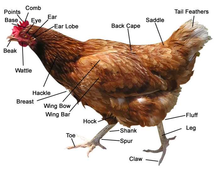
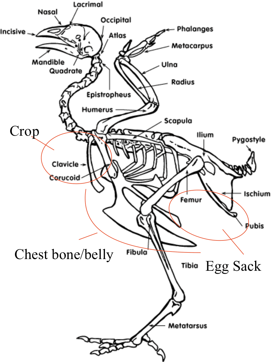
Chickens – Digestive System
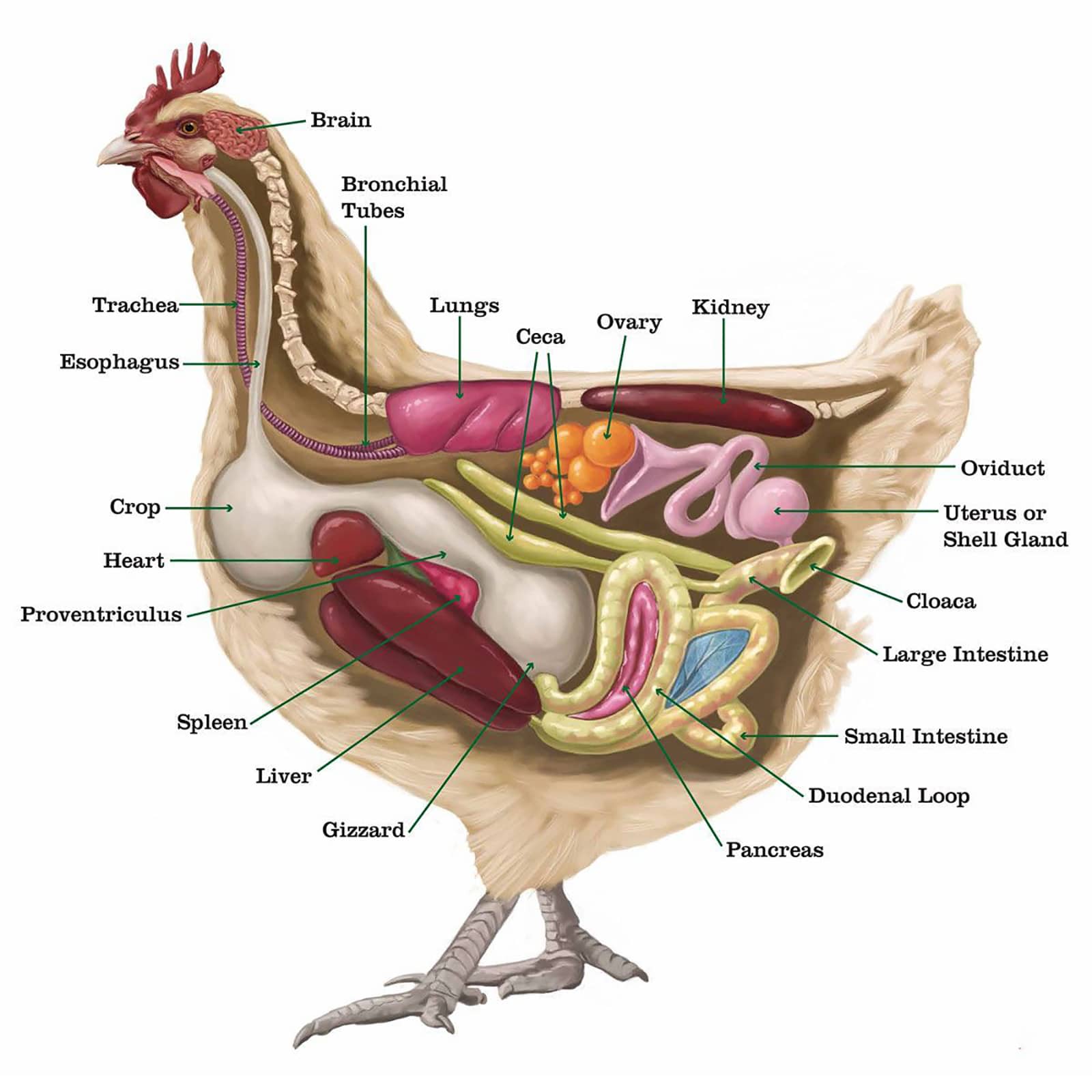
Chickens, as with most birds, use their beaks to pick up food and as they have no teeth they are unable to chew their food. A chicken’s mouth contains glands that secrete saliva which wets the food to make it easier to swallow. The saliva also contains some enzymes which start the digestion process. The chicken’s tongue is then used to push the food to the back of the mouth so that it can be swallowed.
Read more from prestigequeen:
Esophagus
The food then moves into the esophagus which is the flexible tube that connects the mouth with the rest of the digestive tract. It carries food from the mouth to the crop.
The Crop
The crop is an out-pocketing of the esophagus and is located just outside the body cavity in the neck region. Any swallowed food and water is stored in the crop until it is time to pass it on to the rest of the digestive tract. When the crop is empty, or nearly empty, it sends hunger signals to the brain so that the chicken will eat more. Although salivary glands of the mouth secrete the digestive enzyme amylase very little digestion actually takes place in the crop, it is simply a temporary storage pouch.
The crop evolved for birds (in fact it was present in some dinosaurs) that were easy prey for larger animals but which needed to move into the open to find food. This enables them to consume relatively large amounts of food quickly and then move to a more secure location to digest the food they consumed. Occasionally the crop becomes impacted or ‘backed up’ (crop impaction, also referred to as crop binding or pendulous crop).
The Proventriculus
The esophagus continues past the crop to connect the crop to the proventriculus. The proventriculus (also known as the ‘true stomach’) is the glandular stomach where digestion begins. As with human stomachs, hydrochloric acid and digestive enzymes (e.g. pepsin) are added to the food here and digestion begins. At this point, however the food has not yet been ground up. The term ‘proventriculus’ is used since it comes before the ‘ventriculus’ or gizzard, with ‘pro’ being the Latin term meaning before.
The Gizzard
The gizzard, or ventriculus, is a part of the digestive tract unique to birds. It is often referred to as the ‘mechanical stomach’. It is made up of two sets of strong muscles which act as the bird’s teeth. Consumed food and the digestive juices from the salivary glands and the proventriculus pass into the gizzard for grinding, mixing, and mashing, which is aided by the small stones or grit the bird consumes. These stones remain in the gizzard until they become ground into pieces small enough to pass through to the rest of the digestive tract. The stones/grit are weakened by the acidic environment created in the proventriculus and then are ground into tiny pieces by the strong muscles of the gizzard.
Small intestine
The small intestine starts at the exit of the gizzard with the duodenal loop and ends at the ileo-caecal-colon junction. Food in the duodenum is neutralised by the addition of more enzymes excreted by the pancreas. These enzymes break down protein. Also added here is bile produced by the liver and stored in the gall bladder, bile aids in fat digestion. The products of digestion are absorbed from the small intestine and carried to the liver primarily for remanufacture into body tissue or to provide energy.
Caeca
The caeca are two blind-ended tubes which provide space for fermentation. Here undigested food particles are subjected to microbial breakdown. The caeca normally contain a mustard to dark-brown froth which is excreted about once every day.
Large intestine
This part of the gut is very small in the chicken and serves as a further absorption site especially for water.
Cloaca
The cloaca or vent is a chamber common to the digestive and urogenital systems. It is responsible for the elimination of faeces, urine and the passage of eggs or seminal fluid.
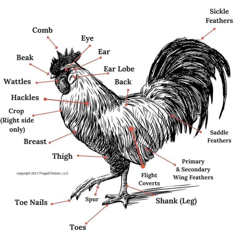
Chicken combs – types and purpose
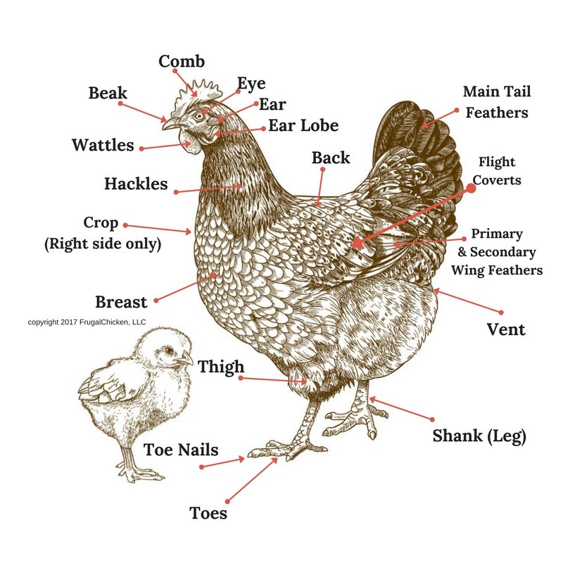
course larger on the cockerel than the hen chicken. There are several forms or shapes of combs including buttercup, cushion, pea, rose, single, strawberry and v-shaped.
Combs are often a distinguishing characteristic that helps identify various breeds and varieties of chickens. In some breeds such as Leghorns and Rhode Island Reds, there are both single comb and rose comb varieties. In addition, the colour of the combs vary from bright red to purple, again depending upon the breed.
The scientific classification of a chicken is Gallus domesticus, where the Latin word ‘gallus’, means comb. So what is the purpose of the chicken’s comb? There appears to be two main reasons. The first is that the combs act as a cooling mechanism for the birds. Chickens cannot sweat to cool themselves, instead, birds are cooled by the blood flowing through the comb and the wattle, which are both externally situated. As the warm blood circulates through the comb, it is cooled and returns to the interior parts of the body, thereby acting as a cooling mechanism in hot weather. This obviously has an adverse effect in cold weather when it is necessary to take precautions to ensure that your chickens do not get frost bite in those areas.
The second reason is that the large combs on males attract females as chickens can detect colour and are very attracted to the colour red.
At one time, many of the commercial poultry farms routinely removed the combs from all birds at an early age to prevent injuries and subsequent infection later in life, which would reduce the commercial value of the bird. Thankfully this is rarely practiced now.
The comb also serves as an indicator of the bird’s health. If it appears lighter or darker than usual or seems to be shriveled or lopped, it is usually a sign of illness. Certainly, it is a sign of whether a bird is in ‘good condition’ when observed at a show. The shape and color of the comb carry a total of 5 points out of 100 in a judge’s evaluation. In addition, a bright red comb on a developing young female (pullet) normally means that the bird is ready to begin her laying cycle.
Types of Chicken Combs
Single combs
The single comb is by far the most common of the comb types and the one that people think of when they think of a chicken’s head. The single comb is a moderately thin, fleshy formation of smooth soft surface texture, firmly attached from the beak along the top of the skull with a strong base. The top portion shows five or six rather deep serrations or distinct points, the middle points being higher than the back or front, forming a semi-oval shape when viewed from the side. The comb is always upright and much larger and thicker in males than in females. It may be lopped or upright in the female. This depends on the breed. The comb is divided into three sections: the front, the middle and that extending past the rear base of the skull, the posterior or blade.
One of the major problems with single combs is that the points tend to freeze and fall off in extremely cold weather. This does not normally affect the health of the bird but does drastically reduce their value as an exhibition bird. Many exhibitors protect their birds by covering the comb with petroleum jelly during times of extreme cold. The petroleum jelly insulates the comb and prevents freezing/frostbite. This is also something that a lot of chicken keepers do as a matter of course to protect their birds in extreme weather even if they are not exhibitors
The males of some breeds of chickens such as the Old English, Modern, and American Games, are required to be ‘dubbed’ in order to be shown. These are all single comb breeds. Dubbing consists of the removal of the head appendages such as comb, wattles, and ear lobes. This is similar to the docking of tails on certain breeds of dogs. This procedure is usually conducted using surgical shears and takes place when the males are six months of age or older. These appendages do not grow back so it is only necessary to perform the procedure once on each bird.
Rose combs
The rose comb is a solid, broad and nearly flat comb on top. It is a low, fleshly comb that concludes in a well-developed tapering spike at the back. It may turn upward as in Hamburg breeds, be nearly horizontal as in Rose Comb Leghorn breeds, or follow the contour of the head as in Wyandotte breeds. The top surface of the main part should be slightly convex and studded with small rounded protuberances. The general shape varies in the different breeds.
Strawberry combs
The Strawberry is a low comb that is set well-forward. The shape and surface resemble the outer part of half a strawberry with the large end nearest to the beak of the chicken.
Cushion combs
The cushion comb is a low, compact chicken comb of relatively small size, it should be quite smooth, possess no depressions or no spikes and not extend beyond the mid point of the skull.
Buttercup combs
The buttercup comb consists of a single blade arising at the juncture of the chicken’s head and beak, rising up and slightly back to the cup shaped crown, set squarely on the centre of the skull. The rim of the cup should bear an evenly spaced circle of points and be closed at the back. Points emerging from the centre of the cup are considered to be a serious defect.
Pea comb
The pea comb is a medium length, low comb, the top of which is marked with three low lengthwise ridges, the centre one being slightly higher than the outer ones, the top of which are either undulated or marked with small rounded serrations. This is a chicken breed characteristic found in Ameraucanas, Brahmas, Buckeyes, Cornish, Cubalayas and Sumatras.
V Shaped comb
The v shaped comb is formed of two well defined, hornlike sections joined at their base, as in Houdans, Polish, Crevecoeurs, La Fleche and Sultans.
Thus a chicken’s comb serves a myriad of purposes from an indicator of health and vitality to a cooling agent to an attraction to the opposite sex. Finally, it seems to add aesthetic value to the overall appearance of the chicken and a big, bright red comb signals that a bird is indeed the ‘cock of the walk’.
The Making of an Egg

An egg is formed gradually over a period of about 25 hours, with many organs and systems helping to convert raw materials from the food eaten by the bird into the various substances that become part of the egg.
The ovary
The hen, unlike most animals, has only one functional ovary, the left one, situated in the body cavity near the backbone. At the time of hatching, a female chick has up to 4000 tiny ova (reproductive cells), from some of which full-sized yolks may develop when the hen matures. Each yolk (ovum) is enclosed in a thin-walled sac, or follicle, attached to the ovary. This sac is richly supplied with blood.
The oviduct
The mature yolk is released when the sac ruptures, and is received by the funnel of the left oviduct (the right oviduct is not functional). The left oviduct is a coiled or folded tube about 80 cm in length. It is divided into five distinct sections, each with a specific function.
Funnel (infundibulum)
The funnel receives the yolk from the ovary. If live sperm are present, fertilisation occurs here. The egg spends about 15 minutes here.
Magnum
The egg spends about three hours here where the inner and outer shell membranes are added, as is some water and mineral salts.
Isthmus
The egg spends about an hour in the isthmus where the albumen (white) is secreted and layered around the yolk.
Uterus or Shell Gland
The egg spends about 21 hours here where initially some water is added, making the outer white thinner. Then the shell material (mainly calcium carbonate) is added and pigments may also be added here, for example, to make the egg brown.
Cloaca (Vagina)
The egg passes through this section before laying.
Poultry Egg Fertilisation and First Hours of Development
Fertilisation of the ova (yolk) by the sperm takes place in the infundibulum about 15 minutes after its holding sac ruptures and releases it. The combination of the ova and the sperm form a single cell called the zygote. The zygote reaches the isthmus about 5 hours after fertilisation and it is here that the embryo starts to develop, through cell division. By the time it leaves the isthmus it has grown to an eight cell embryo and by the time it leaves the uterus it has grown to 256 cells. The developing embryo will stay at this stage until incubation conditions are right for further development.
Cell division continues as the egg passes along the oviduct and is laid. An egg takes about 25 hours from the release of the ova to the point of lay. This means that in actual fact a chicken egg takes 22 days to hatch, not the 21 days which is usually quoted: 1 day in the oviduct and 21 days in the nest or incubator.
Formation of the ectoderm, endoderm and mesoderm
As cell division continues two layers are formed over the yolk these are the ectoderm (uppermost) and the endoderm (underneath) layers. At about this stage the central cells of the blastoderm ( the layer of cells that develops on the surface of the yolk and gives rise to the germinal disk from which the embryo develops) separate from their contact with the yolk and form a cavity. It is in this cavity that subsequent embryo development occurs. Soon after the formation of the ectoderm and endoderm, a third layer of cells called the mesoderm or middle layer is formed.
The important organs and body tissues develop from these three layers. With the ectoderm producing the nervous system, parts of the eyes, the feathers, beak, claws, and skin. The endoderm produces the respiratory system, the digestive system, and secretory organs. While the mesoderm produces the skeleton, muscles, circulatory system, reproductive organs, and excretory system. the segregation of cells into different tissue functions occurs before the egg is laid.
Extra Embryonic Membranes
Not only do the early embryonic germ layers (ectoderm, mesoderm, and endoderm) develop into specialised tissues of the body, but also they form membranes outside the body which help protect and nourish the developing chick embryo. Three extra-embryonic membranes are formed from the these first germ layers and are as follows:
The Yolk Sac
The yolk sac envelops the yolk and draws nourishment from it, producing an enzyme that changes the yolk material to a form that can be used as a food source by the developing embryo. Any remaining, unused yolk material in the yolk sac when the chicken hatches from the egg are drawn into the abdomen for use by the chicken for the first two to three days after hatching, while the chicken learns what to eat and drink and where to find it.
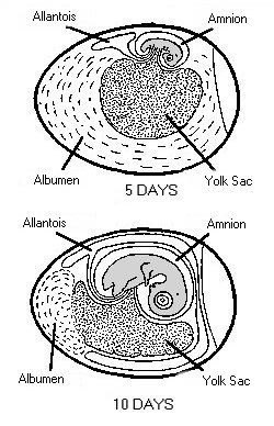
The Amnion
Ectoderm and mesoderm surrounding the embryo, form a protective covering over the embryo called the amnion. The inner layer of cells secretes amniotic fluid in which the embryo floats. This fluid keeps the embryo from drying out and helps absorb shocks when the egg is knocked.
The Chorio-Allantoic Membrane
The chorion which lines the eggshell and performs gas exchange and waste elimination is formed from the ectoderm and mesoderm layer and the allantois is the embryo’s connection to the chorion. The allantois appears as a balloon-like structure coming from the hindgut at about 4 or 5 days. The allantois develops an extensive circulatory system connected to that of the embryo and driven by the new embryonic heart. When the allantois is fully developed it completely surrounds the embryo.
The allantois membrane has a number of functions, including a respiratory role. The growing embryo uses oxygen and produces carbon dioxide, but is unable to carry out respiration itself yet and therefore it is the role of the allantois to oxygenate the blood and eliminate the carbon dioxide.
The allantois serves an excretory role, removing waste that the embryo metabolizes. It also has a digestive role, providing a means for the embryo to access the albumen and the calcium of the shell.
Read more from prestigequeen:
- Beginner’s Guide to Raising Backyard Chickens
- Chicken Illnesses, Disease and Injuries
- Everything you ever wanted to know about Eggs
Contents